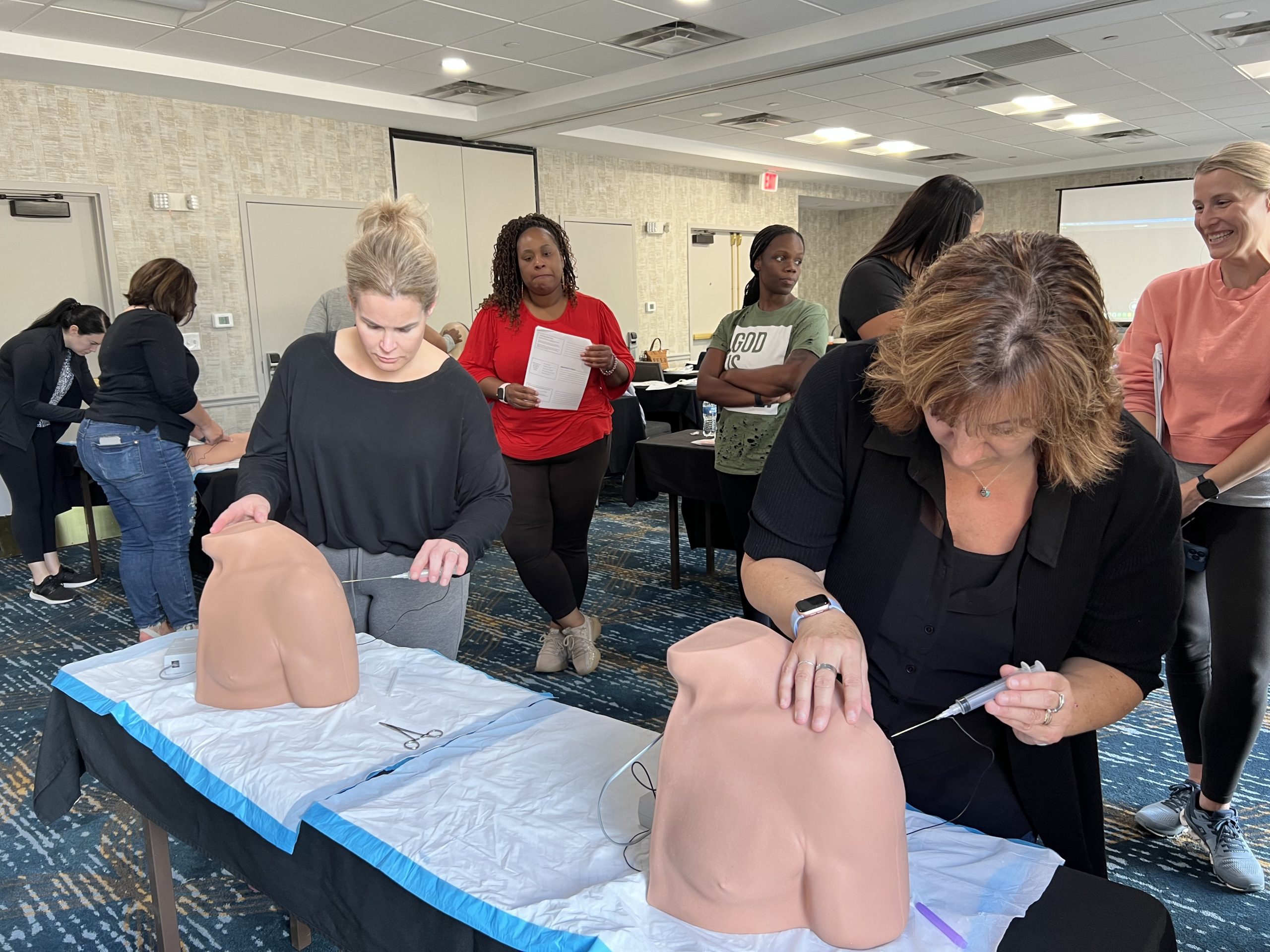
What is Suturing?
Suturing is the preferred method of wound closure to achieve hemostasis in clean, uncontaminated wounds. Use correct techniques, such as direct pressure, hemostats, and sutures to minimize bleeding risks; an anesthetic agent to provide pain control; and a clean field for laceration repair to decrease contamination.
Explore the wound for foreign bodies and debris carefully, because one-third of foreign bodies could be missed on initial inspection. Consider the use of diagnostics methods such as X-rays, ultrasonography, or computed tomography.
Assess tendons, nerves, joints, muscles, and vessels for tissue damage. Immediate orthopedic referral is necessary for severe crush wounds, severely contaminated wounds, open fractures, tendon and muscle lacerations, nerve damage, laceration involving eyes, eyelids, and salivary ducts, or injuries that require sedation for repair.
Avoid the use of povidone-iodine solution, hydrogen peroxide, and detergents for wound irrigation, due to possible damage to fibroblasts that could delay wound healing.
Why?
To provide hemostasis, decrease risk of infection, and promote wound healing.
How?
Pre-procedure steps
Supplies:
- Suture kit (non-absorbable suture material, forceps, needle driver)
- Sterile gloves/clean gloves
- Suturing
- Normal saline, sterile water, or potable tap water
- Anesthetics (lidocaine, epinephrine, tetracaine)
- Antiseptic agent (Hibiclens®, chlorhexidine, betadine)
- Syringes and needle for anesthetics
Suture Materials
There are two types of sutures: absorbable and non-absorbable.
Absorbable sutures are used for closing deep, multiple layer lacerations. Types of absorbable sutures include polyglactin 910 (Vicryl®) and poliglecaprone (Monocryl®). They typically absorb within 4 to 8 weeks.
Non-absorbable sutures are used for tissue repair, but they must be removed. They include nylon and monofilament (polypropylene [Prolene®]). Silk sutures are not recommended for wound closure due to high tissue reactivity and poor tensile strength.
In practice, a 3-0 or 4-0 suture is recommended for the trunk, 4-0 or 5-0 for scalp and upper and lower extremities, and 5-0 or 6-0 on the face.
Consider the use of absorbable 3-0 or 4-0 sutures for mucosal lacerations, which include tongue, genitalia, and mouth) for better cosmetic outcomes.
Common Suture Techniques in Urgent and Primary Care Setting
Simple interrupted technique
What?
The simple interrupted suture technique is commonly used and is effective for general wound closure. This technique provides good cosmesis because the wound is everted and retracts as it heals.
Why?
To provide wound closure, decrease infection, and promote wound healing. The technique allows for some portion of the sutures to be removed and others left in, if wound is irregular or becomes infected.
How?
Pre-procedure Steps
- Radiographs should be performed, if a fracture or foreign body is suspected.
- Wound irrigation should be performed for contaminated wounds (dirt or debris) using a 16- to 19-gauge catheter attached to a 35-60mL syringe.
- Determine tetanus prophylaxis and update as needed.
Supplies:
- Suture kit (choice of suture material, forceps, needle driver)
- Sterile gloves/clean gloves
- Suturing
- Normal saline or potable tap water (for wound irrigation)
- Anesthetics (lidocaine, epinephrine, tetracaine)
- Antiseptic agent (Hibiclens®, chlorhexidine, betadine)
- Syringes (35-60mL) for anesthetics
- Needle (25- to 27- gauge) for anesthetics
- 4 X 4 cotton gauze
- Tape
Procedure Steps
Proper Technique for an Instrument Tie
- Grasp the needle side of the suture with the non-dominant hand.
- Pull suture partway through, leaving a 1-inch tail (Step 1).
- Point needle holder at tail of suture.
- Use the non-dominant hand to throw 2 loops of suture around the end of the needle holder (Step 2).
- Grasp the tail suture end with the needle holder and pull through the 2 loops (Step 3).
- Throw 1 loop of suture around the end of the needle holder (Step 5).
- Tie the knot and trim the ends (Step 6).

Figure 3.1 Proper technique of an instrument tie.
Simple Interrupted Suture
- Infiltrate tissue with local anesthesia, injecting the needle around the wound bed and surrounding area, using a fanlike approach.
- Pull back the plunger to aspirate and avoid blood vessels.
- Cleanse the wound with an antiseptic agent.
- Apply the needle to the needle driver.
- Clasp needle 1/2 to 2/3 away from tip.
- Penetrate the wound edge with the needle driver, 1/4-inch bite away from the wound edge at 90 degrees.
- Reach into the wound and grasp the needle with the needle driver, taking equal bites.
- Pull it free to give enough suture material to enter the opposite side of the wound.
- Pull suture partway through, leaving a 1-inch tail.
- Point needle holder at tail of suture.
- Use the left hand to throw 2 loops of suture around the end of the needle holder.
- Grasp the tail suture end with the needle holder and pull through the 2 loops.
- Throw 1 loop of suture around the end of the needle holder.
- Tie the knot and trim the ends of the suture.
- Each suture is cut before another one is started.
- Insert the needle perpendicular to the epidermis.
- Traverse the epidermis and the full thickness of the dermis.
- Exit perpendicular to the epidermis on the opposite side of the wound.
- Tie suture off and trim leftover.
- Repeat technique until completed.
- The two sides of the stitch should be symmetrically placed in terms of depth and width (Figure 3.2).

Figure 3.2 Simple Interrupted Suture.
Post-Procedure Steps
- Apply cotton gauze and tape to the sutured area.
Clinical Checkpoints
- Consider timing of closure:
- Wounds on the face and scalp can be closed up to 24 hours post injury.
- Trunk and extremity wounds can be closed within an 8- to 12-hour window.
- To decrease acidity, warm the local anesthesia to body temperature by buffering it with sodium bicarbonate.
- Recheck laceration repair within 48 hours to reduce infection risk and ensure proper wound healing in patients with diabetes mellitus, immunocompromised patients, and those on long-term corticosteroids.
- Refer patients to general surgery if they present with a deep dermal laceration, penetrating wounds, extreme contamination, or wounds with nerve involvement.